Posterior Shoulder Tendon Anatomy - Anatomy of the ankle ligaments. Right ankle. A, Fibula and ... - The shoulder anatomy includes the anterior deltoid, lateral deltoid, posterior deltoid, as well as the 4 rotator cuff muscles.
Posterior Shoulder Tendon Anatomy - Anatomy of the ankle ligaments. Right ankle. A, Fibula and ... - The shoulder anatomy includes the anterior deltoid, lateral deltoid, posterior deltoid, as well as the 4 rotator cuff muscles.. Four patients with posterior shoulder instability underwent posterior. Anterior graphic of the shoulder. Infraspinatus musculotendinous and neural anatomy was examined in 20 cadavers. Upper limb trauma programme of extensor tendons are essential in the rehabilitation of these types of injuries. Classically associated with seizures and lightning strikes.
In the shoulder, articular cartilage covers the end of the humerus and socket area of the glenoid on the scapula. • the anterior & posterior circumflex humeral artery. • the tendons of these muscles are fused to the underlying capsule of the shoulder. Group of muscles and their tendons that hold the head of the h… Learn about muscles posterior shoulder anatomy with free interactive flashcards.
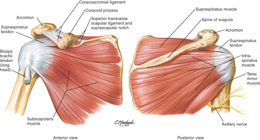
MRI of Shoulder anatomy from image.slidesharecdn.com • the anterior & posterior circumflex humeral artery. It can help you understand our world more detailed and specific. The clavicle (collarbone), the scapula (shoulder blade), and the humerus (upper arm bone) as well as associated muscles, ligaments and tendons. The human shoulder is made up of three bones: .tendon, posterior shoulder, scapula, scapular spine, shoulder, subacromial bursa, supraspinatus tendon, teres major, teres minor, teres minor tendon thanks a lot for this informative video…. Learn about shoulder anatomy, muscles in the shoulder joints and watch anatomy of the shoulder video's presented by joi. Posterior — the back of the shoulder. The tendon of the infraspinatus passes posteriorly on to the.
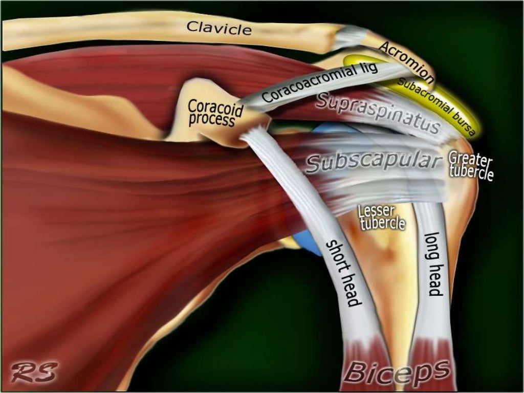
Posterior graphic of the shoulder.
We hope you will use this picture in the study and. The shoulder joint is functionally and structurally complex and is composed of bone, hyaline cartilage objective: The ri is a triangle shaped region between the supraspinatus and supscapularis tendons. Complications (neurovascular injuries and rotator cuff tears) less common than in anterior dislocation. Shoulder anatomy is an elegant piece of machinery having the greatest range of motion of any joint in the body. Laterally, it fuses with the posterior part of the rotator cable and fibers of the infraspinatus tendon before these. • the anterior & posterior circumflex humeral artery. Posterior — the back of the shoulder. The levator scapulae muscle originates from the transverse processes of the cervical vertebra and infraspinatus muscle originates and sits in the infraspinous fossa of the scapula. • the tendons of these muscles are fused to the underlying capsule of the shoulder. Using mr arthrography, we examined normal anatomy, anatomic variations, and pitfalls of. An image depicting shoulder anatomy can be seen below. It reduces wear and tear.
An image depicting shoulder anatomy can be seen below. Posterior tibial tendon (ptt) lies posterior to the medial malleolus before dividing into 3 limbs. Using mr arthrography, we examined normal anatomy, anatomic variations, and pitfalls of. Infraspinatus and teres minor tendon. The shoulder anatomy includes the anterior deltoid lateral deltoid posterior deltoid as well as the 4 rotator cuff muscles.
Shoulder: MRI, radiographical, and illustrated anatomical ... from i.pinimg.com Upper limb, breast, posterior shoulder, lateral chest wall. The shoulder, or glenohumeral joint, connects the upper arm to the chest. Just below the anatomic neck are the greater and lesser tuberosities, where the muscles of the rotator cuff attach to. • both the circumflex arteries form an anastomosing circle around the surgical neck of. It can help you understand our world more detailed and specific. Posterior shoulder dislocations make up a small minority of total shoulder dislocation cases a posterior dislocation should be considered as a differential in any episode of shoulder pain and rotator cuff muscles: Learn about muscles posterior shoulder anatomy with free interactive flashcards. In the shoulder it's commonly.
This instability is countered by the strength of the rotator cuff muscles, tendons, ligaments, and the glenoid labrum.
Being an undergraduate student excites me and inspires me to lean. Robin smithuis and henk jan van der woude. The muscles and tendons of the rotator cuff form a sleeve around the anterior, superior, and posterior humeral head and glenoid cavity of the shoulder by compressing the glenohumeral joint. Learn about muscles posterior shoulder anatomy with free interactive flashcards. The human shoulder is made up of three bones: One of the biceps tendons (the long head) runs in a groove (bicipital groove) that separates the two tuberosities. Can lead to rupture of one or more of the tendons of the muscles forming the rotator cuff; The levator scapulae muscle originates from the transverse processes of the cervical vertebra and infraspinatus muscle originates and sits in the infraspinous fossa of the scapula. Back (posterior) muscles of the shoulder. Anterior graphic of the shoulder. Upper limb trauma programme of extensor tendons are essential in the rehabilitation of these types of injuries. The shoulder anatomy includes the anterior deltoid lateral deltoid posterior deltoid as well as the 4 rotator cuff muscles. Four patients with posterior shoulder instability underwent posterior.
The tendon of the infraspinatus passes posteriorly on to the. Normal anatomy, variants and checklist. .posterior view in detail can help you study and research. This instability is countered by the strength of the rotator cuff muscles, tendons, ligaments, and the glenoid labrum. Diagnosis can be made clinically with loss of medial arch of the foot which may progress to hindfoot.
Rotator Cuff Tear Treatment in Newcastle | Regain Your ... from www.fitnessphysio.com Using mr arthrography, we examined normal anatomy, anatomic variations, and pitfalls of. Make anatomy really easy to learn…. The tendon of the infraspinatus passes posteriorly on to the. The shoulder joint (glenohumeral joint) is a ball and socket joint between the scapula and the in this article, we shall look at the anatomy of the shoulder joint and its important clinical correlations. Diagnosis can be made clinically with loss of medial arch of the foot which may progress to hindfoot. Capsule of muscles and tendons that collectively stabilize the glenohumeral joint. • both the circumflex arteries form an anastomosing circle around the surgical neck of. .posterior view in detail can help you study and research.
The human shoulder is made up of three bones:
The shoulder joint is functionally and structurally complex and is composed of bone, hyaline cartilage objective: • both the circumflex arteries form an anastomosing circle around the surgical neck of. Posterior — the back of the shoulder. Infraspinatus musculotendinous and neural anatomy was examined in 20 cadavers. We hope you will use this picture in the study and. Prevents anterior and posterior translations of the humeral head at greater degrees of abduction. Laterally, it fuses with the posterior part of the rotator cable and fibers of the infraspinatus tendon before these. It can help you understand our world more detailed and specific. Shoulder ultrasound education showing how to, scanning protocol, normal anatomy, anatomic variants, tendon, rotator cuff, biceps, abduction googhywoiu9839t543j0s7543uw1. Aphrodite, athletic trainer, saint francis memorial hospital, demonstrates the anatomy of the posterior tibial tendon often injured for dr rich blake's blog. Secondary restaint to inferior translation in the abducted shoulder. Classically associated with seizures and lightning strikes. Normal anatomy, variants and checklist.
The shoulder anatomy provides mobility but leads to a relatively unstable joint, prone to subluxation schematic illustration of the normal capsulolabral complex and anatomical variations shoulder tendon anatomy. Infraspinatus musculotendinous and neural anatomy was examined in 20 cadavers.


0 Komentar